Bookface Sinus Model
Aids strong informed consent, protecting both patient and surgeon in sinus surgery
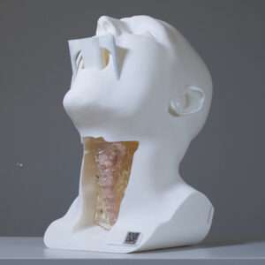
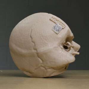
The Anatomex 3D Bookface Sinus Model is a unique anatomical communication and education tool designed primarily for ENT consultations, helping patients visualise and understand their sinus pathology and treatment options. This in turn leads to improved patient consent, protecting both the patient and the surgeon. It also serves as an invaluable training model for general sinonasal education.
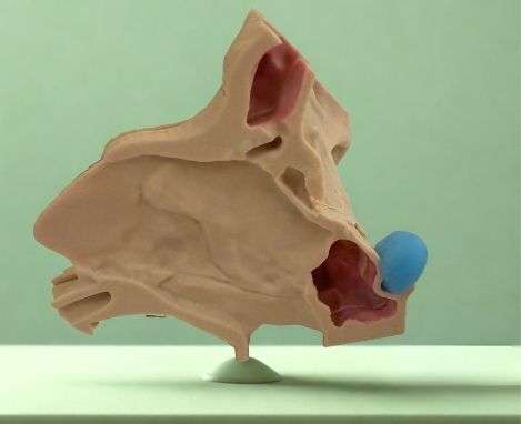
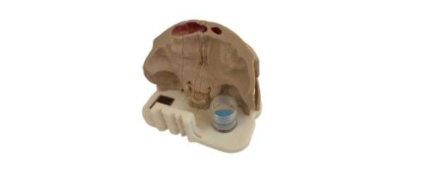
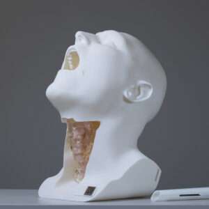
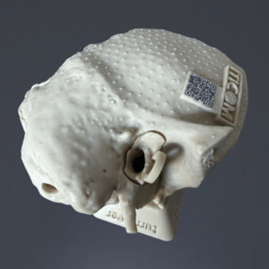
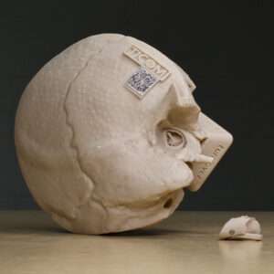
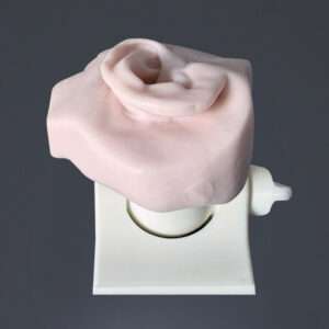
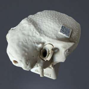
This life-size anatomical representation of the midface accurately depicts the frontal, ethmoid, maxillary, and sphenoid sinuses. Its innovative, sliced ‘book-style’ design allows the model to open, providing a clear, multi-angled view of intricate sinus structures. For easy identification, the turbinates are coloured blue, the ethmoid cells purple, and the remaining paranasal sinuses pink. One side of the nasal septum is also covered with mucosa to assist in demonstrating septoplasty procedures.
Developed in collaboration with Professor Darlene Lubbe—a global leader in endoscopic sinus, transorbital, and skull base surgery—the model combines advanced engineering and materials for exceptional anatomical accuracy and lifelike realism. It is an effective tool for improving patient understanding and informed consent, as well as an engaging educational and display piece for any medical or training setting.
Key features of the Bookface Sinus Model
- Clear identification of the frontal, ethmoid, maxillary, and sphenoid sinuses when the midface is open.
- Distinctly coloured sinuses for effortless recognition.
- Light-colored base material for enhanced aesthetics, making it an ideal display piece for a physician’s desk.
- Anatomically accurate design, ensuring precision in surgical training and patient education.
- Clearly visible anatomical structures, including the nasal septum with mucosa on one side to assist in demonstrating septoplasty surgery.
- Small and compact, a perfect fit for any desk, measuring just 13L x 13W x 9.5H (cm)
An indispensable tool for teaching and patient education
I’ve been very pleased with the Bookface sinus model since I started using it. It has greatly improved how I explain sinus anatomy and procedures to both patients and students. The layered design makes it easy to demonstrate the 3D structure of the sinuses, the role of the turbinates in nasal airflow, and the impact of septal deviation. It has been especially helpful when discussing endoscopic sinus surgery, septoplasty, and septal perforation repair, as patients can clearly visualise the anatomy and surgical steps.
I also find the option to place the supplied putty-like adhesive in different sinuses valuable for illustrating tumours, foreign bodies, and skull base surgery. Overall, it has made explanations much clearer and I highly recommend it for both clinical and teaching settings.
Professor Hesham Saleh, leading international figure in rhinoplasty, London, UK
All anatomy included in the model
Sphenoid Bone
Ethmoid Bone
Lacrimal Bone
Nasal Bone
Zygomatic Bone
Maxilla
Maxillary Sinus
Sphenoid Sinus
Sphenoid Sinus Ostium
Frontal Sinus
Superior Turbinate
Middle Turbinate
Inferior Turbinate
Lamina Papyracea
Orbital Plate
Agger Nasi Cell
SLICE 1
All anatomy except Septum
Agger Nasi CellUncinate Process
Ethmoid Bulla
Anterior Ethmoid Cells
Posterior Ethmoid Cells
SLICE 2
Septum
Frontal Sinus
Uncinate Process
Ethmoid Bulla
Anterior Ethmoid Cells
Posterior Ethmoid Cells
Basal Lamella of the Middle Turbinate
Infraorbital Nerve
Infraorbital Foramen
Posterior Ethmoidal Artery
Anterior Ethmoidal Artery
Sphenopalatine Artery Foramen
Optic Nerve Canal
Crista Galli
Cribriform Plate
Olfactory Fossa
SLICE 3
Superior Turbinate
Middle Turbinate
Inferior Turbinate
Lamina Papyracea
Sphenoid Sinus Ostium
Sphenoid Sinus
SLICE 4
Maxillary Sinus
Infraorbital Nerve
Included in the Bookface Sinus Model kit
- 3D Bookface Sinus Model – with hand-painted sinus structures for easy anatomical identification.
- A5 laminated whiteboard — double-sided, featuring model segments, septum, and midface scan. The whiteboard provides ENTs with a practical teaching aid for illustrating surgical approaches, marking incision lines, and demonstrating procedures such as septoplasty or sinus access.
- Whiteboard marker with built-in eraser.
- Anatomex pen
- Perspex pointer – ideal for indicating structures on the model while keeping it clean (avoiding ink marks from pens).
- Carry bag – provides a convenient and protective way to transport the model on ward rounds, to consulting rooms, or between training venues
- Tumour demonstration box – includes modelling putty for simulating obstructions or tumour placement.
- Care and maintenance instructions — printed inside the lid of the box for quick reference.
Makes Explaining Treatment Options Much Easier
Using the Bookface sinus model has made my consultations much easier – patients instantly grasp what’s going on inside their nose and sinuses. It helps turn complex anatomy into something very tangible, which makes discussions about treatment options far more effective.
Shahz Ahmed, Consultant Rhinologist & Skull Base Surgeon, University Hospitals Birmingham, UK


Shahz Ahmed –
Makes Explaining Treatment Options Much Easier
Using the Bookface sinus model has made my consultations much easier – patients instantly grasp what’s going on inside their nose and sinuses. It helps turn complex anatomy into something very tangible, which makes discussions about treatment options far more effective.
Professor Hesham Saleh –
An Indispensable Tool for Teaching and Patient Education
I’ve been very pleased with the Bookface sinus model since I started using it. It has greatly improved how I explain sinus anatomy and procedures to both patients and students. The layered design makes it easy to demonstrate the 3D structure of the sinuses, the role of the turbinates in nasal airflow, and the impact of septal deviation. It has been especially helpful when discussing endoscopic sinus surgery, septoplasty, and septal perforation repair, as patients can clearly visualise the anatomy and surgical steps. I also find the option to place the supplied putty-like adhesive in different sinuses valuable for illustrating tumours, foreign bodies, and skull base surgery. Overall, it has made explanations much clearer and I highly recommend it for both clinical and teaching settings.
Ola Basson –
When it wasn’t on my desk anymore, I suddenly realised how much I depended on it.
Werner Rossouw (verified owner) –
Brilliant concept to help patients better understand their operations, which is not also great for insight but directly translates into better outcomes for me and my patients as they are then much more inclined to stick to post-operative care protocols! A win-win in many ways!