
 Transorbital Surgery Book


Air-41 Airway Model
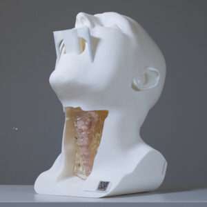
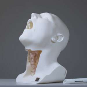
A modular, anatomically accurate 3D simulation model designed for hands-on airway training. It supports rigid and flexible bronchoscopy, suspension laryngoscopy and supraglottic airway insertion. Interchangeable tracheal sections simulate different levels of stenosis, allowing trainees to practise balloon dilatation and other airway interventions in a controlled training environment.
Introducing the Air-41 Airway Model
The Air-41 Airway Model is a high-fidelity, modular 3D training model engineered for advanced airway management simulation. Designed in collaboration with leading ENT specialists, it provides a highly realistic platform for teaching, demonstrating, and practising a comprehensive range of airway access methods.
It replicates the nasal cavity, oral cavity, oropharynx, hypopharynx, larynx, and proximal trachea in accurate anatomical proportions. The model is designed for hands-on training in endoscopic airway assessment, balloon dilation procedures, and laryngoscopy technique
Supported procedures and interventions
The Air-41 airway model allows trainees to develop the fine motor skills required for safe and effective airway management. Supported techniques include:
- Rigid and flexible bronchoscopy
- Suspension laryngoscopy
- Supraglottic airway (LMA) insertion
- Balloon dilation
- Stent placement
Flexible modular design
To replicate true-to-life conditions, the model features visible ventilation via inflatable balloons and simulates tracheal stenosis at multiple levels. Its versatile, modular design allows instructors to easily swap replaceable tracheal sections to recreate different pathological scenarios. This enables trainees to fully understand the complexities of airway obstruction and confidently execute a variety of interventions.
Uses for the Air-41 Airway Model
Lightweight and durable, the model is ideal for:
- Training workshops and medical simulation centres
- Skills assessment and competency-based learning
- Product demonstrations for airway devices and instruments
With its blend of anatomical realism, modular flexibility, and educational value, the Air-41 Airway Model brings airway training to a new level of precision and engagement.
Optional add-on: Eustachian Tube Soft Tissue Insert
The Air-41 is available with an optional upgraded soft tissue insert that includes anatomically accurate bilateral Eustachian tube (ET) orifices. This add-on is designed for institutions and companies wishing to expand their simulation curriculum to include Eustachian tube dilation training alongside the standard tracheal airway training scenarios.
About the Eustachian Tube Insert
The ET insert is a direct replacement for the standard soft tissue section. It preserves all the features of the standard insert — nasal passages, oral cavity, oropharynx, and larynx — while adding bilateral Eustachian tube orifices within the nasopharynx. The orifices are dimensioned to accommodate commercially available ET balloon dilators and to allow confident scope navigation to the tubal opening.
- Anatomically accurate bilateral ET orifices positioned within the nasopharynx
- Compatible with 0° and 30° rigid Hopkins rod telescopes for direct visualisation
- Suitable depth and calibre to accommodate standard ET balloon dilators
- All standard nasal, oral, and laryngeal anatomy retained from the standard insert
- Inserted and removed using the same technique as the standard soft tissue section (see Section 2)
Supported training scenarios
With the ET insert in place, the following training scenarios can be conducted:
- Endoscopic identification and localisation of the Eustachian tube orifice via transnasal rigid endoscopy
- Navigation of a balloon dilator catheter to the ET orifice under direct 0° or 30° Hopkins rod vision
- Balloon Eustachian tuboplasty (BET) technique — catheter advancement, positioning, and staged inflation
- Graded balloon inflation under continuous endoscopic monitoring
- Bilateral ET dilation training in a single session
- Combined ET and airway simulation using both scopes in sequence
Below: Testing the Air-41 Airway Model using a Karl Storz endoscope and a DISA Medinotec Trachealator non-occlusive airway dilatation balloon inserted through a suspension laryngoscope mouthpiece supplied with the model.

Above: Karl Storz endoscope inserted into the trachea (note the visible glow); preparing to insert the Trachealator non-occlusive airway dilatation balloon.
Related products


Rhinology
An anatomically accurate 3D simulation model designed for hands-on endonasal surgical training, including functional endoscopic sinus surgery (FESS). Its removable, replaceable cartridges replicate relevant sinonasal anatomy and enable repeated practice of surgical techniques. Additional cartridges can be purchased separately, providing a flexible, cost-effective training solution for surgeons and surgical trainees.
Products
A portable, reusable surgical simulation platform designed for hands-on training in transoral and endoscopic laryngeal procedures. Its integrated suspension system, transparent laryngeal anatomy and interchangeable pathology inserts support training in endoscope navigation, instrument positioning and procedural techniques using rigid and flexible endoscopes, lasers, coblation systems and powered instruments.


Rhinology
US$240 | £180 | €210 A life-size anatomical model designed to help ENT surgeons explain sinus anatomy, pathology and treatment options to patients. Its opening, book-style design and colour-coded structures provide clear views of the paranasal sinuses, making it easier to demonstrate findings from nasal endoscopy and CT scans, as well as proposed surgical procedures.


Transorbital surgery
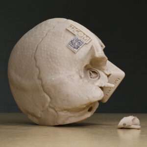
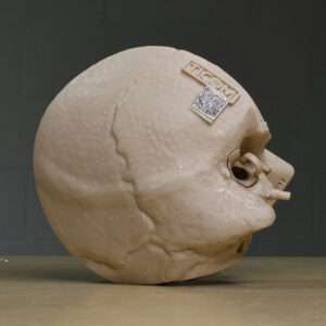
A life-size, anatomically accurate 3D model designed for hands-on transorbital endoscopic and skull base surgical training. Its interchangeable orbital components allow surgeons to explore complex orbital and skull base anatomy, practise surgical approaches and simulate procedures involving the sphenoid sinus and middle cranial fossa. Transparent and solid orbital modules, including options with integrated soft tissue and Sternberg canal defects, support anatomical teaching and practical surgical simulation.
Otology
An anatomically detailed simulation model representing the temporal bone of a 64-year-old adult, designed for hands-on otologic surgical training. The Temporal Bone MAX supports procedures ranging from middle ear surgery to mastoidectomy and cochlear implantation, depending on the configuration. Features include realistic bone-drilling characteristics and anatomical structures relevant to advanced ear surgery.