




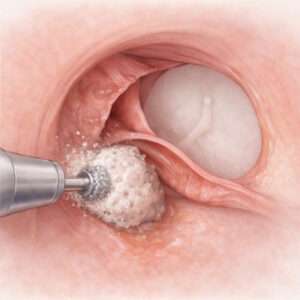
Exostosis Model
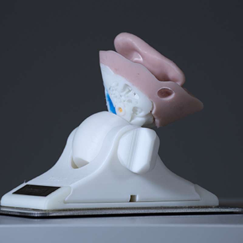
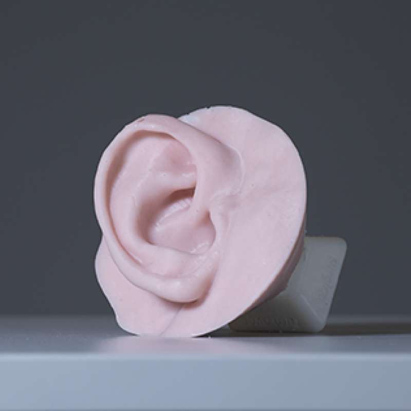
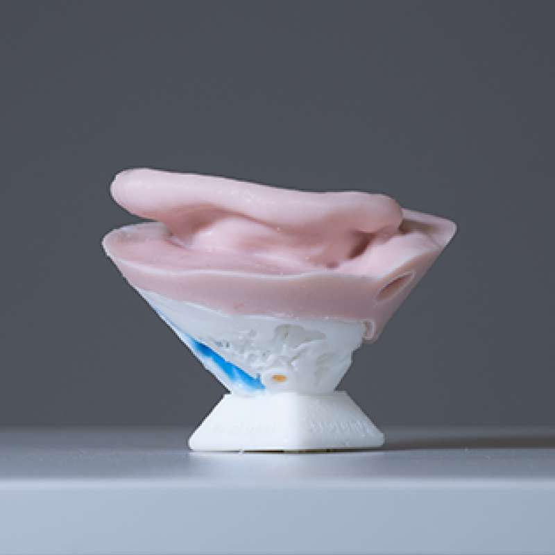
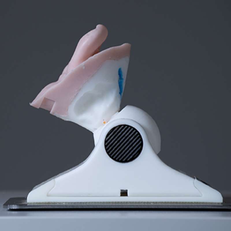
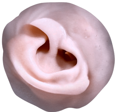
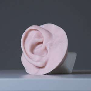
The Exostosis Model (EXO-41) is a high-fidelity surgical simulation platform specifically engineered for drill-based canalplasty and exostosis surgery training. Featuring anatomically accurate bony exostoses and integrated soft tissue, this model provides realistic tactile and haptic feedback for powered instrumentation, hammer-and-chisel techniques, and endoscopic visualisation. With colour-coded critical structures – including the facial nerve and chorda tympani – it reinforces procedural safety and nerve preservation, offering a modular, cost-effective, and repeatable training solution for residency programs and specialised otology workshops.
Exostosis model: High-fidelity simulation for drill-based canalplasty
The Exostosis Model (EXO-41) is a high-fidelity surgical simulation platform engineered for exostosis surgery training, with a focus on powered instrumentation and drill-based canalplasty. Designed for surgical skills courses and residency programs, this model provides a realistic environment to refine operative technique, hand positioning, drill control, and procedural safety.
Anatomical precision and material realism
Our Exostosis Model replicates the external auditory canal (EAC) with anatomically accurate dimensions and representative bony exostoses.
Advanced composite materials deliver:
- Realistic tactile feedback: Skin incision resistance and bone removal response using powered drills or hammer-and-chisel techniques.
- Auditory and haptic accuracy: True-to-life drilling feedback for enhanced procedural confidence.
- Critical structure integration: Anatomically positioned facial nerve and chorda tympani reinforce safe surgical technique and nerve preservation.
Anatomical structures included
- External auditory canal with bony exostosis
- Ear canal skin (1 mm thickness)
- Tympanic membrane
- Color-coded facial nerve for easy identification
- Chorda tympani
- Color-coded sigmoid sinus
- Representative mastoid air cells
Supported surgical techniques
Using standard otologic instrumentation, the Exostosis Model supports:
- Endoscopic, Endaural, and Post-Auricular Approaches
- Canalplasty and Exostosis Surgery
- Tympanoplasty
- Ventilation Tube (Grommet) Insertion
Optimised for powered instrumentation training
The model is specifically designed for drill-based workflows, enabling:
- Continuous drilling and bone removal
- Precise instrument angulation within a narrow ear canal
- Controlled tissue handling under microscopic or endoscopic visualisation
Modular and cost-effective architecture
The Anatomex system is built for scalability and long-term value. Each Exostosis Model is supplied with:
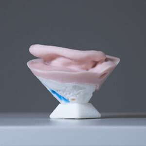
- Stable mounting: Choice of an A6 metal plate or thick A4 perspex base with an optional raised rubber rim for fluid containment.
- Rotatable mount: Flexible positioning to match surgical scenarios.
- Self-supporting design: Stands independently on a desk for pre- and post-dissection patient demonstrations.
Key system advantages
- Reusable infrastructure: Base plates and mounting systems are compatible with all Anatomex3D surgical models and retained by your facility.
- Replaceable surgical segments: Only procedure-specific components require replacement, reducing long-term cost per delegate.
- Scalable training: Ideal for recurring surgical education courses and high-volume training programs.
Summary
The Exostosis Model is the definitive training platform for surgeons requiring realistic, repeatable simulation of exostosis surgery and canalplasty. It builds technical skill, procedural confidence, and surgical safety in a controlled, high-fidelity environment – without the ethical and logistical challenges of cadaveric specimens.
Related products


Otology
The Temporal Bone Model 64-Year-Old is a high-fidelity surgical simulation tool designed for advanced otology and neurotology procedures, offering ultra-realistic anatomical detail and surgical feedback. Available in three configurations (MINI, MIDI, MAX), this model enables surgeons to practice procedures like cochlear implantation, mastoidectomy, and ossiculoplasty, with features such as realistic bone drilling feedback, a functional ossicular chain, and a hollow cochlea for implant simulations, ensuring a precise and repeatable training experience.


Otology
The Temporal Bone Model 2-Year-Old is a high-precision synthetic otology model designed to replicate real surgical conditions. It features anatomically accurate structures, including the facial nerve, cochlea, and ossicles, allowing for hands-on training in procedures such as cochlear implantation, mastoidectomy, and ossiculoplasty. With lifelike bone texture, a self-draining system, and a patented removable tympanic membrane, this model enhances surgical precision and confidence.


Otology
The Temporal Bone Ear Model is a high-fidelity otologic training platform designed for advanced temporal bone surgery, featuring a realistic external "flesh" ear for authentic anatomical orientation. This model provides ultra-realistic tactile and acoustic drilling feedback, supporting complex procedures such as mastoidectomy, cochlear implantation, and facial nerve decompression. With integrated features like fluid-filled structures for immediate feedback on surgical safety and a modular, cost-effective design, it offers a precise, repeatable, and ethical alternative to cadaveric specimens for residency programs and advanced surgical workshops.